A body system is a collection of organs that work together to perform a specific task.
The 11 body systems in the human body which work together to ensure the body has sufficient nutrition and oxygen, can expel toxins and is able to deal effectively with pathogens and disease.
Each of the 11 systems rely on each other to work together in harmony to be able to perform their function. When all the body systems are healthy, the person is able to maintain a state of homeostasis, by which the internal conditions necessary to maintain life remain stable and constant.
 Select to learn more
Select the items on the image to reveal the key organs and main functions of each body system.
Image by OlgaChernyak, Shutterstock, Shutterstock licence
Source: "How bodies work" in Overview of Body Systems by Lumen Learning
Now let’s explore each system in more detail.
Cardiovascular system
The cardiovascular system (also called the circulatory system) is made up of the heart and the blood vessels leading to and from the heart.
The heart has four chambers and is made up of muscular walls and four valves.
The cardiovascular system is responsible for transporting blood, hormones and nutrients such as glucose to all parts of the body. It works in conjunction with the respiratory system to provide oxygen to cells, tissues and organs.
 Watch the video
Watch ‘Amoeba sisters biology learning playlist’ to learn more about the body’s circulatory system and the path that blood follows as it flows around the body.
Then complete the quiz questions.
 Check your understanding
Respond to the questions below and select ‘Check’ to see if you are correct. Select the 'Next' button to move to the next question, then select 'Check' again for feedback.
 Important
Veins are
blood vessels that bring
oxygen-depleted blood towards the heart (with the exception of the pulmonary
vein, which carries oxygenated blood).
Arteries are blood vessels that take oxygen-enriched blood away from the heart (with the exception of the pulmonary artery, which carries deoxygenated blood to the lungs).
Components of the cardiovascular system
The cardiovascular system consists of the heart, the blood vessels and the blood.
The heart is located slightly to the left of the middle chest (thorax) region and has four chambers: two ventricles and two atria.
The left side of the heart consists of a top chamber (atria) and a lower chamber (ventricle) this is repeated on the right side of the heart. The atria and the ventricles are separated by valves that open and close in time with the heartbeat.
There is a dividing layer of tissue running lengthwise through the middle of the heart called the septum.
The blood vessels of the body consist of arteries, veins and capillaries. The major artery of the body is the aorta and is shaped like a candy cane. Arteries are very strong and elastic and take blood away from the heart.
The major vein in the body is the vena cava, which transports blood back to the heart. Veins are less elastic than arteries and contain small flaps, called valves, that assist with the transportation of blood back to the heart by stopping blood from flowing backwards.
Capillaries are the smallest blood vessels of the body.
Capillary walls are only one cell thick, which allows substances to move through the capillary wall and into the cells of the surrounding tissue. The exchange of oxygen and carbon dioxide happens at this level. Capillaries eventually join the arterial and venous systems of the blood vessels at the ‘capillary bed’.
 Select to learn more
Select the items on the image to reveal specific terminology relating to the structures of the cardiovascular system.
Image by brgfx, Freepik, Licence
Select to learn more
Select each bar to expand and reveal further information about the cardiovascular system.
 Watch the video
Watch ‘What is blood made of? What does blood do?’ to learn more about the role and make up of blood.
Then complete the quiz questions.
Check your understanding
Respond to the questions below and select ‘Check’ to see if you are correct. Select the 'Next' button to move to the next question, then select 'Check' again for feedback.
Respiratory system
The respiratory system is located in the head, neck and chest cavity. The main function of the respiratory system is to provide oxygen to the cells of the body and allow for the release of waste carbon dioxide from the body in the exhalation.
The respiratory system consists of the lungs, diaphragm, bronchi, trachea, larynx, pharynx and nose.
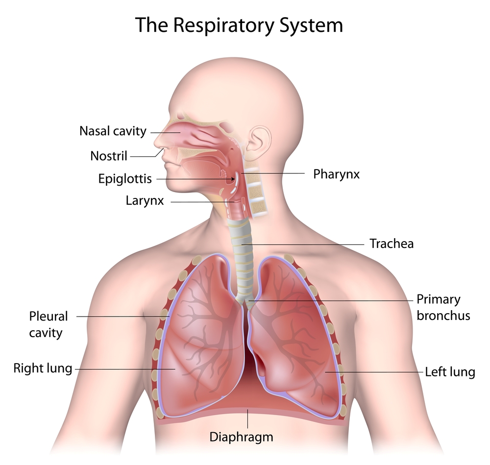
Image by Alila Medical Media, Shutterstock, Shutterstock licence
Air is breathed in through our nose and sometimes mouth. The air is warmed and any unwanted material is caught in the mucous of the nose and expelled from the body when we blow our nose.
The air then enters the oropharynx, which is a shared passageway for food and air.
A little flap of tissue called the epiglottis closes the airway when we swallow, stopping foods and liquids from entering the trachea. The air continues through the larynx, where the vocal cords are located and then travels through the trachea, before it enters the lungs.
Important
The lungs are two large hollow organs located in the chest cavity.
They are divided into lobes and are protected by the rib cage and various muscles.
The air touches the surface of the lungs where oxygen transfuses through the alveoli (small grapelike structures at the end of the bronchioles) and across the capillary wall into the blood. At the same time, carbon dioxide leaves the capillary to be expelled from the lungs.
Any particles of dust or pollen that enter the lungs are trapped in the mucous that is secreted by the goblet cells of the lungs and swept in an upward fashion by cilia (small hair like structures that line the respiratory passage) to be expelled by the body through coughing or sneezing.
The lungs also have specialised cells called macrophages that engulf foreign material and thus protect the body.
 Watch the video
Watch ‘Respiratory system – Basic anatomy’ to learn more about the respiratory system.
Then answer each of these questions:
- Name the organs in the upper and lower respiratory zones.
- Why is the trachea made of cartilage rings?
- Where does gas exchange occur?
- Describe the process of gas exchange.
Musculoskeletal system
The musculoskeletal system incorporates two major systems: the muscular system and the skeletal system.
The muscular system is made up of almost 700 muscles. It holds us upright and governs movement and posture and moves blood and other substances through the body.
The skeletal system comprises the 206 bones of the human body from the skull to the toes.
The skeleton plays a number of roles including:
- bones protect important organs, such as the skull protects the brain and the rib cage protects the lungs
- other bones help a person stay upright
- the skeleton acts as a frame for the body’s muscles, other tissues and skin
- the skeleton also makes movement possible.
Important
The skeletal muscles work with the skeletal system to:
- enable ambulation (walking) and other movement
- support the upright posture
- regulate the flow of food from the mouth to the anal sphincter
- control breathing by changing pressure in the lungs
- regulate temperature
- enable nonverbal communication – facial muscles are used to smile, frown, create rapport and show disapproval.
Terminology of the skeletal system
The terminology used to describe the skeletal system includes the structures that make up the human body and the classification of bones.
- Bones provide the form and structure of the body and protect organs such as the brain, lungs and heart.
- Axial skeleton forms the head and main trunk of the body and consists of the skull, spinal column (vertebrae), ribs and sternum.
- Appendicular skeleton forms the extremities of the body and consists of shoulder girdle, arm bones, pelvic girdle and leg bones.
- Long bones are the hard, dense bones that provide structural strength and mobility, such as the femur.
- Ligaments are bands of tough, fibrous connective tissue that connect bones to other bones at joints or connect bones to other structures such as cartilage. They provide stability and support to the joints.
- Tendons are flexible cords of strong fibrous collagen tissue that attach muscle to bone. They transmit force from muscle contractions to the bones, enabling movement.
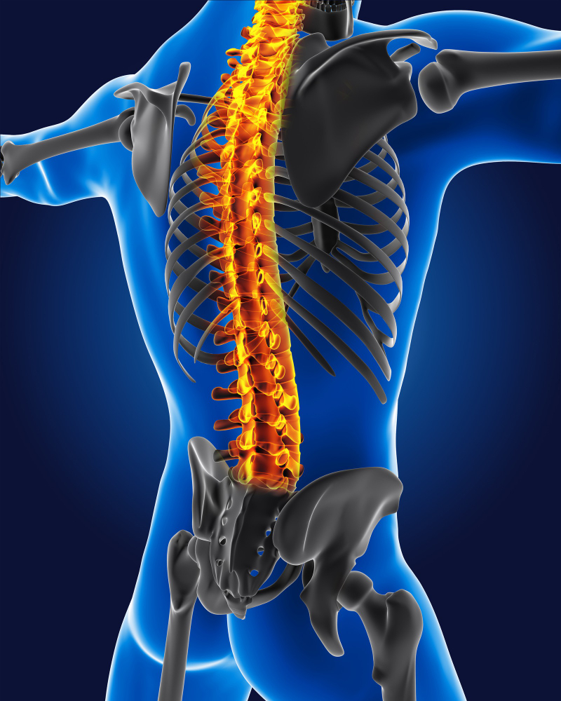
Image by kjpargeter, Freepik, Licence
Our bones, which provide the form and structure of our bodies, are broken down into the following.
Select to learn more
Select the items on the image to reveal more information about each type of bone.
Terminology of the muscular system
Terminology is broken down into the structures that support the muscular system and the movements our bodies make.
Select to learn more
Select each bar to expand and reveal further information about muscles and movements.
 Watch the video
Watch ‘How your muscular system works’ to learn more about the musculoskeletal system.
Then complete the quiz questions.
Check your understanding
Respond to the questions below and select ‘Check’ to see if you are correct. Select the 'Next' button to move to the next questions, then select 'Check' again for feedback.
Endocrine system
One of the major functions of the endocrine system is to keep the body in balance.
Homeostasis is the medical term that describes these regulatory processes that keep variables stable and within safe limits (e.g., body temperature and blood glucose level).
One of the systems that plays a major role in homeostasis is the endocrine system.
The endocrine system produces and secretes hormones that are distributed throughout the body and regulate other functions of the body.
Hormones regulate:
- bone density
- growth
- heart rate
- metabolism
- mood
- organ function.
Endocrine glands secrete specific hormones that target other glands, organs of the body or whole body systems. They work to maintain homeostasis. Many of the hormones secreted are vital to body maturation and sexual reproduction and some are essential for vital body functioning.
 Select to learn more
Select the items on the image to reveal more information about the endocrine system.
Image by Designua,
Shutterstock, Shutterstock licence
The function of the endocrine system
The endocrine system is a collection of glands that produce hormones to regulate metabolism, growth, development, tissue function, sexual function, reproduction, sleep and all the functions required for development and everyday life.
- Endocrine glands secrete hormones directly into the bloodstream, which carries them to the target organs or receptor sites.
- A hormone is a chemical message that acts as a ‘key in a lock’ for its target receptor. They carry specific messages and cannot attach to or ‘unlock’ another receptor.
- Hormones and their effects are closely regulated by a system of feedback loops. An example of this is the release of insulin in response to the level of glucose in the blood after a meal.
| Gland | Hormone | Function |
|---|---|---|
| Thyroid | Thyroxine | Regulates metabolism and temperature. |
| Calcitonin | Inhibits the release of calcium from the bones. | |
| Parathyroid | Parathyroid hormone | Stimulates the release of calcium from the bones. |
| Islet cells (in the pancreas) | Insulin | Decreases blood sugar by promoting the uptake of glucose by the cells. |
| Glucagon | Increases blood sugar by stimulating the breakdown of glycogen in the liver. | |
| Testes | Testosterone | Regulates sperm cell production and secondary sex characteristics in males. |
| Ovaries | Oestrogen | Stimulates egg maturation and controls secondary sex characteristics in females. |
| Progesterone | Prepares the uterus to receive a fertilised egg. | |
| Adrenal medulla | Epinephrine (‘fight’ hormone) | Activates the flight or fight response. |
| Norepinephrine (‘flight’ hormone) | ||
| Adrenal cortex | Glucocorticoids | As part of the stress response they increase blood sugar levels and decrease immune response. |
| Aldosterone | Regulates sodium levels in the blood. | |
| Pineal gland | Melatonin | Controls sleep cycles and reproductive cycles. |
 Watch the video
Watch ‘Endocrine system introduction’ to learn more about the endocrine system. Then complete the quiz questions.
Check your understanding
Respond to the questions below and select ‘Check’ to see if you are correct. Select the 'Next' button to move to the next question, then select 'Check' again for feedback.
Digestive system
The digestive system breaks down food and makes its nutrients available to the body. The system then distributes these nutrients and eliminates the waste products of digestion.
The digestive system comprises a long winding tube that includes hollow organs along its path, such as the stomach and intestines, as well as accessory organs such as the liver and gallbladder.
It extends from the mouth (the start of the digestive system) to the anus (the end of the digestive system).
 Select to learn more
Select the items on the image to reveal each component of the digestive system and what function they each serve.
Image by Vecton, Shutterstock, Shutterstock licence
The function of the digestive system
The function of the digestive system is to transform the food we eat into the nutrients the body requires for healthy functioning. That is, it mechanically and chemically processes food and eliminates waste products.
 Read to learn more
Select the arrows to move through the slides and learn about the six essential nutrients humans require.
 Watch and answer
Watch ‘How your digestive system works’ and answer the question as it appears.
Urinary system
The urinary system, also called the excretory system or renal system (renal means kidneys), allows the body to filter the blood and eliminate liquid waste called urea. This system also helps to keep chemicals (such as potassium and sodium) and water in balance.
Urea is produced when foods containing protein (such as meat, poultry and certain vegetables) are broken down in the body. Urea is carried in the blood to the kidneys where it is removed, along with water and other wastes, in the form of urine.
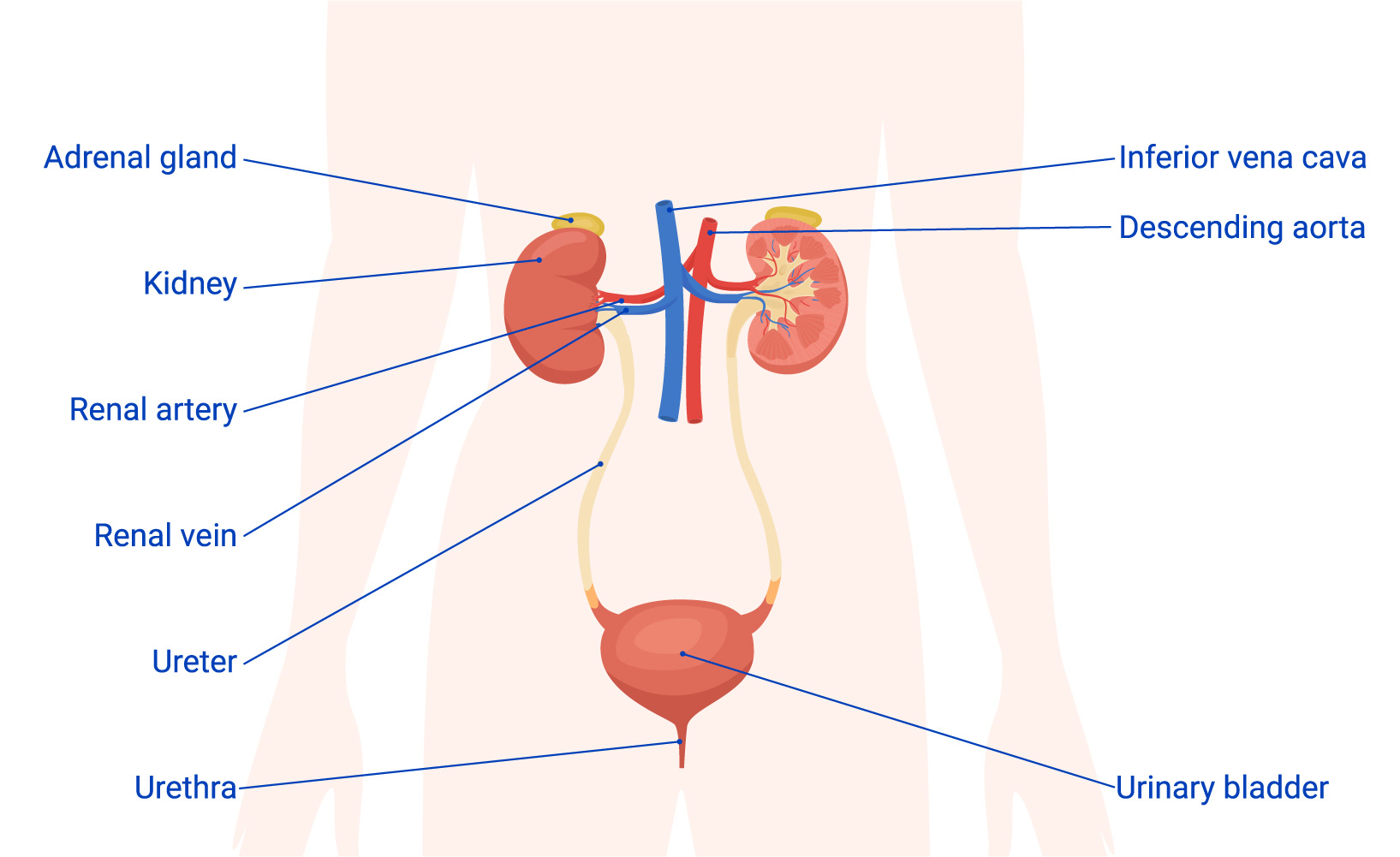
Image by macrovector, Freepik, Licence
Select to learn more
Select each bar to expand and reveal further information about the urinary system.
The kidney contains thousands of small filters called nephrons, which are the functional unit of the kidney and filter around 180 litres of blood per day. Each nephron is made up of a very small filter, called a glomerulus, which is attached to a tubule. As blood passes through the nephron, fluid and waste products are filtered out.
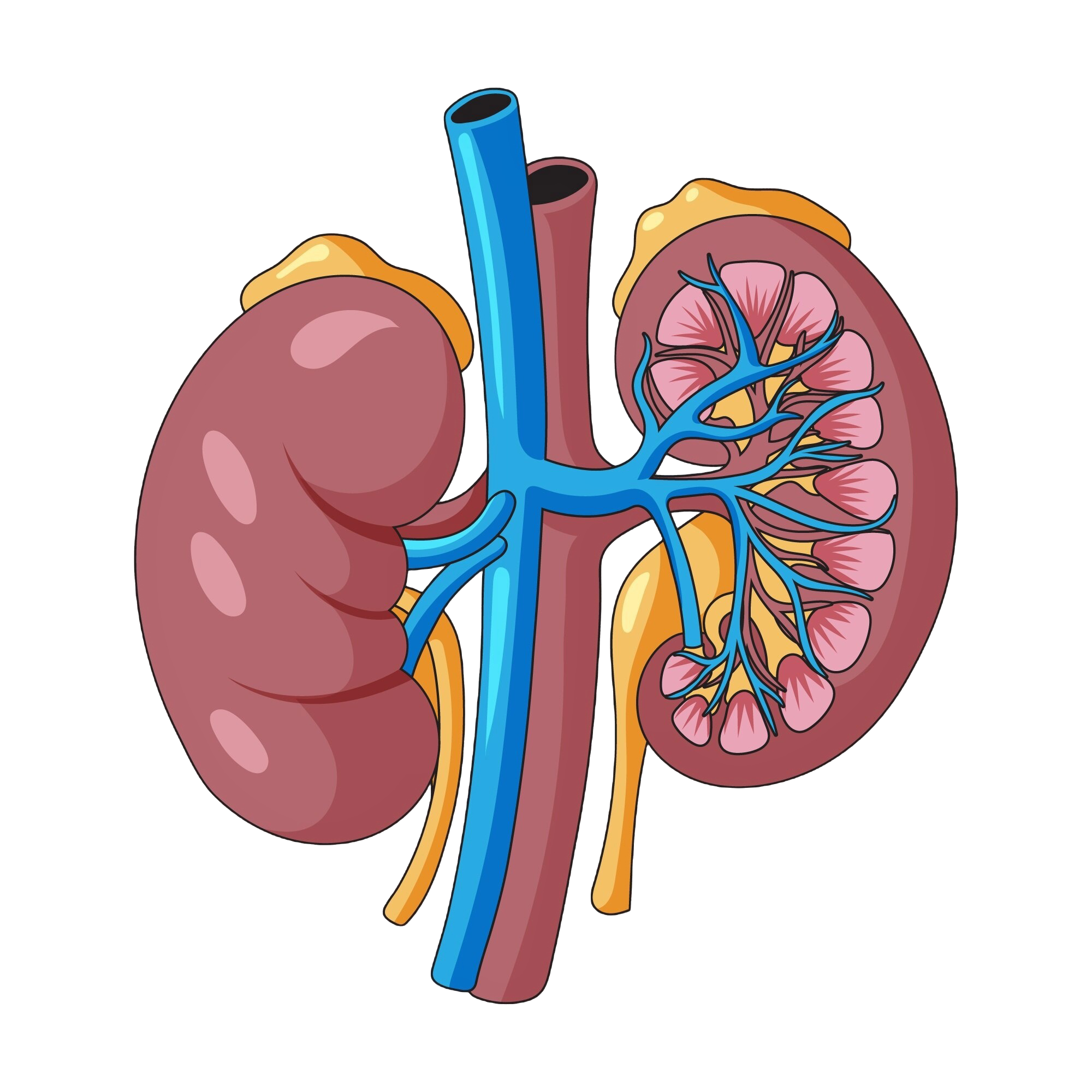
Image by Freepik, Freepik, Licence
Waste products and fluid not required by the body leave in the form of urine. This fluid is transported by the ureters to the bladder.
The bladder is able to expand to hold urine and when it is stretched beyond a certain level, receptor cells in the wall of the bladder send a message to the brain that creates the urge to urinate. The body then expels the urine through the urethra to exit the body.
Any fluid or substance that is required by the body is then reabsorbed by the nephrons and returns to the bloodstream.
Important
- Normal, healthy urine is a pale straw or clear yellow colour.
- Darker yellow or honey-coloured urine often means you need more water.
- A darker, brownish colour may mean a liver problem or severe dehydration.
- Pinkish or red urine may mean blood in the urine.
 Watch the video
Watch ‘How do your kidneys work’ to learn more about the function of the kidneys. Then complete the quiz questions.
Check your understanding
Respond to the questions below and select ‘Check’ to see if you are correct. Select the 'Next' button to move to the next questions, then select 'Check' again for feedback.
Reproductive system
The reproductive system of females and males is the system by which humans develop into sexually mature adults capable of reproducing, that is, having babies.
It controls the development of secondary sex characteristics such as breast development in females and the growth of pubic hair in both males and females.
The female and male reproductive systems differ in structure but have the same overall functions:
- to cause the maturation of the human body (the secondary sex characteristics that develop a boy into a man and a girl into a woman)
- to make reproduction possible – the creation of a new person.
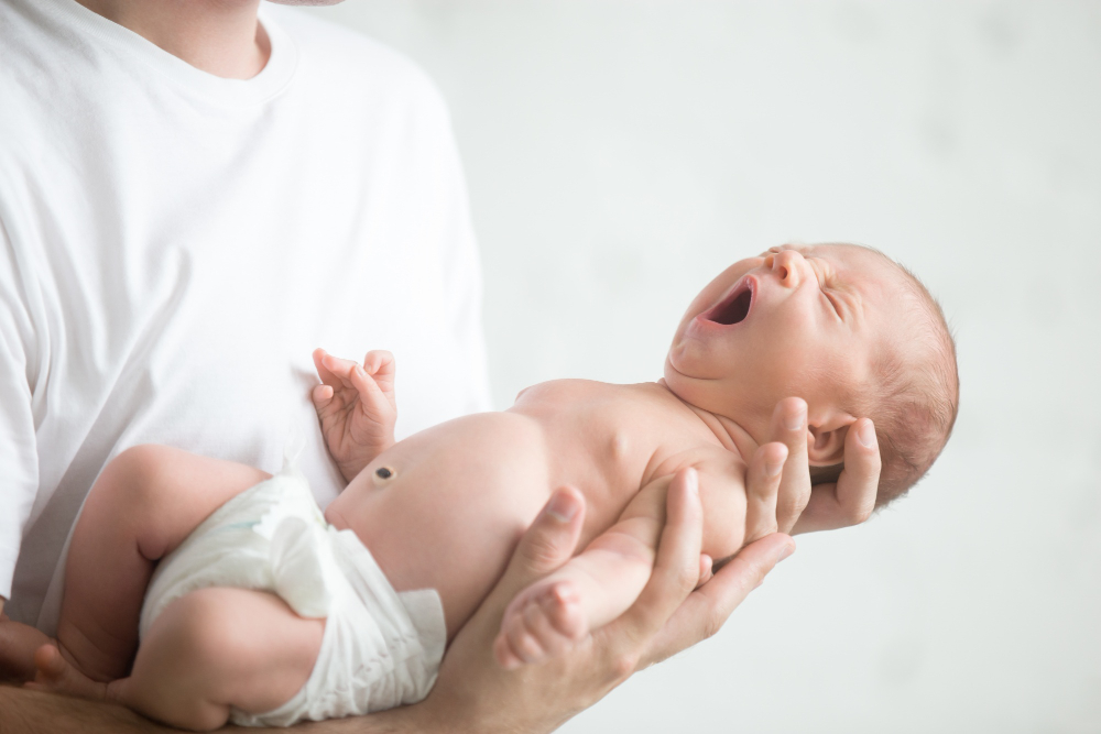
Image by yanalya, Freepik, Licence
Male reproductive system
The main function of the male reproductive system is:
- to produce and transport gametes (sperm) and its protective fluid, semen
- to eject the sperm into the female reproductive tract
- to produce and secrete male reproductive hormones.
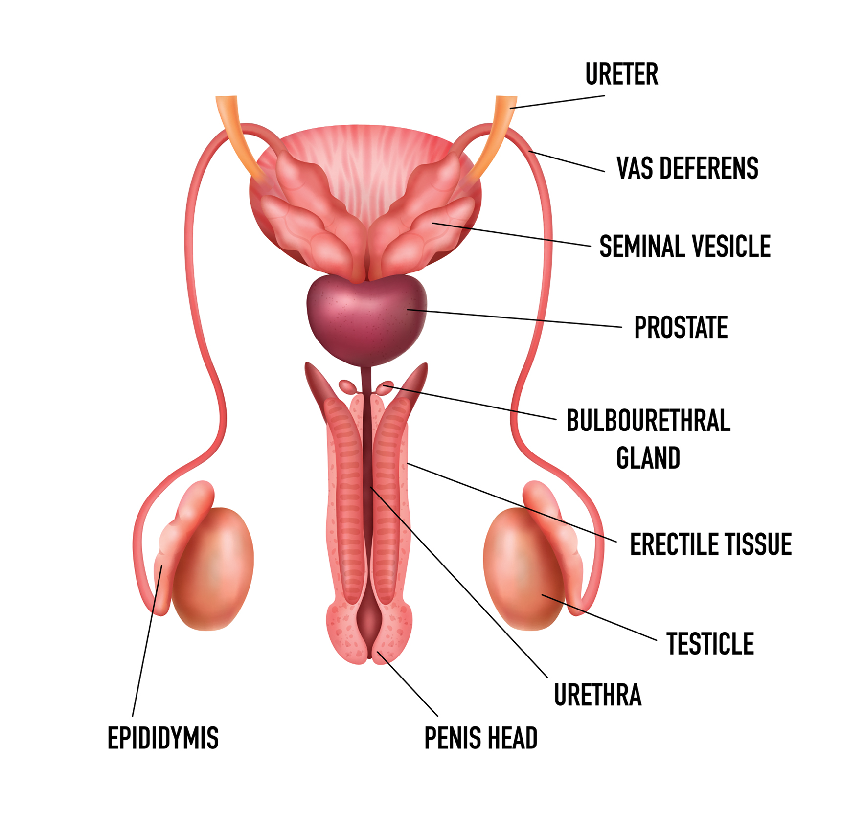
Image by Macrovector, Shutterstock, Shutterstock licence
| Structure Type | Description |
|---|---|
| External structures |
|
| Internal structures |
|
Important
Male hormones
Follicle stimulating hormone is necessary for sperm production, known as spermatogenesis, while luteinising hormone triggers the production of testosterone. This stimulates the development of male characteristics, such as muscle mass, fat distribution, bone mass, facial hair, voice change, development of the sex organs and sex drive.
Female reproductive system
The functions of the female reproductive system include producing gametes (ova, or eggs), secreting sex hormones (such as estrogen), providing a site for fertilisation, gestating a foetus if fertilisation occurs, giving birth to a baby and breastfeeding a baby after birth.
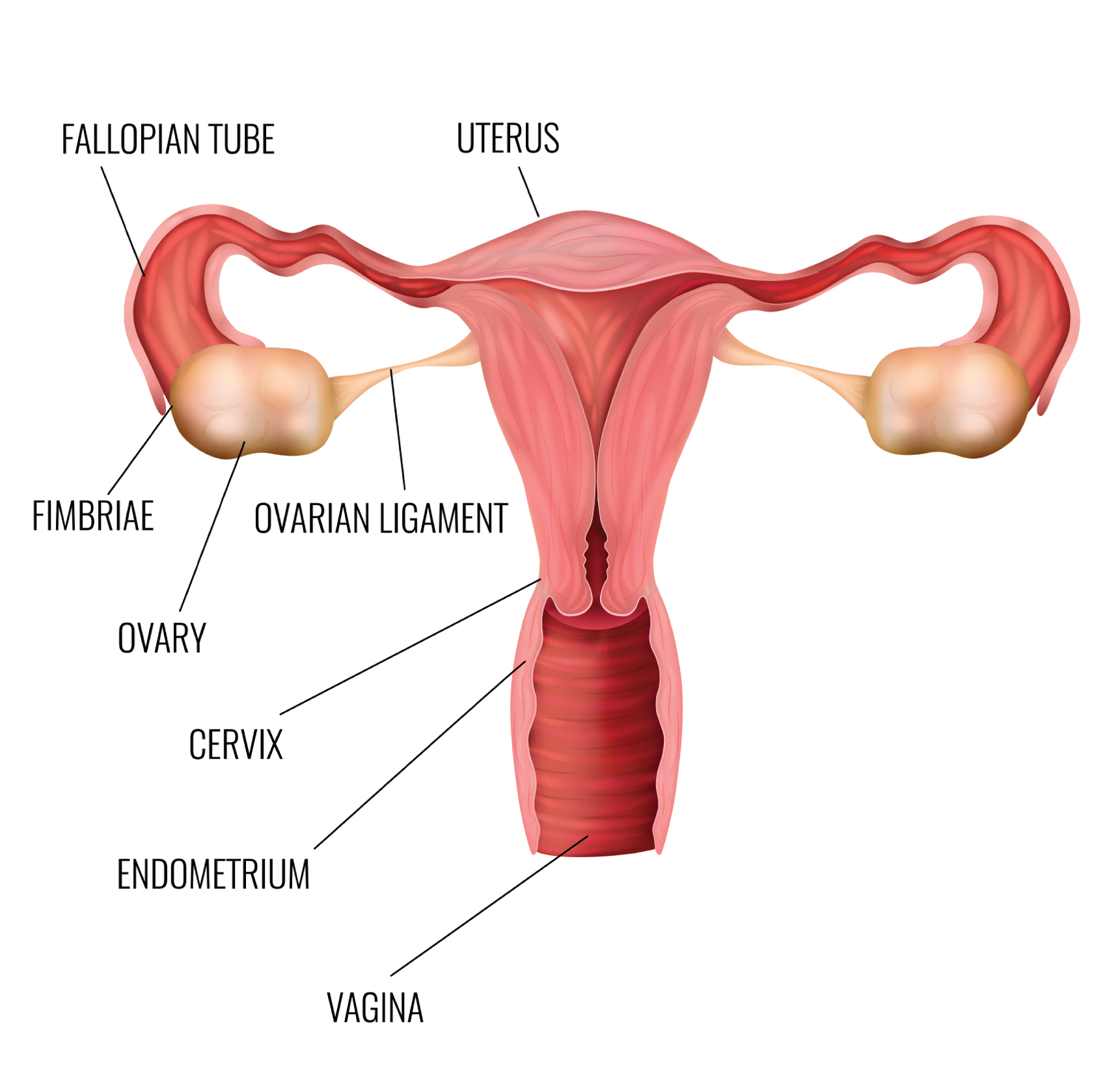
Image by Macrovector, Shutterstock, Shutterstock licence
| Structure Type | Description |
|---|---|
| External structures |
|
| Internal structures |
|
Important
Female hormones
Follicle-stimulating hormone (FSH) prompts the maturation of an egg in the ovary, while Luteinising hormone (LH) triggers the release of the egg.
These hormones originate from the brain and circulate through the blood to the ovaries, stimulating the growth of approximately 15 to 20 eggs in the ovaries. Oestrogen and progesterone, active after puberty, sustain the uterus lining and promote egg maturation in the ovaries, leading to their regular release during the menstrual cycle.
The menstrual cycle
The female menstrual cycle is a natural process controlled by hormones, involving stages of egg maturation, ovulation and shedding of the uterine lining. This cycle has two phases, the follicular phase and the ovarian phase.
The follicular phase commences on the first day of the period. Follicle stimulating hormone and luteinising hormone are circulated to stimulate the ovaries to produce ova. These hormones also stimulate the increase of oestrogen.
The ovarian phase of the menstrual cycle also consists of two phases, the ovulatory phase and the luteal phase. The ovulatory phase commences when oestrogen levels rise, which results in the release of an ovum. This is ovulation. The ovum is then caught by the fimbriae (finger-like projections) at the end of the fallopian tubes. The ovum progresses through the fallopian tube where fertilisation can take place.
After ovulation, the follicle that released the ovum starts secreting progesterone, which prepares the lining of the uterus for implantation of the fertilised ovum. The lining becomes thick and lush. If no fertilisation takes place, this lining breaks down and is expelled from the body during menstruation.
Sexuality
All people have a right to express their sexuality.
As a support worker, you must understand this right and be considerate of how people may express their sexuality differently. This is just as important for older adults and people with disabilities as it is for any other age group and must be addressed with appropriate respect and confidentiality.

Image by frimufilms, Freepik, Licence
You can assist the people you support by:
- respecting their privacy and upholding principles of confidentiality
- speaking to your manager about referring them to an appropriate professional (counsellor, therapist or psychologist) if they express concerns about their attractiveness, sex life or relationships
- ensuring that individuals participate in a full range of social activities that support them to find a partner, should they wish to
- not judging their sexual choices
- respecting the person’s desire to have children (consult with your manager about referring the person to an appropriate healthcare professional).
Check your understanding
Respond to the questions below and select ‘Check’ to see if you are correct. Select the 'Next' button to move to the next question, then select 'Check' again for feedback.
Integumentary system
The integumentary system is made up of the skin, exocrine glands (that secrete substances outside of the body), hair and nails.
This system plays various roles, with the skin serving as its primary organ.
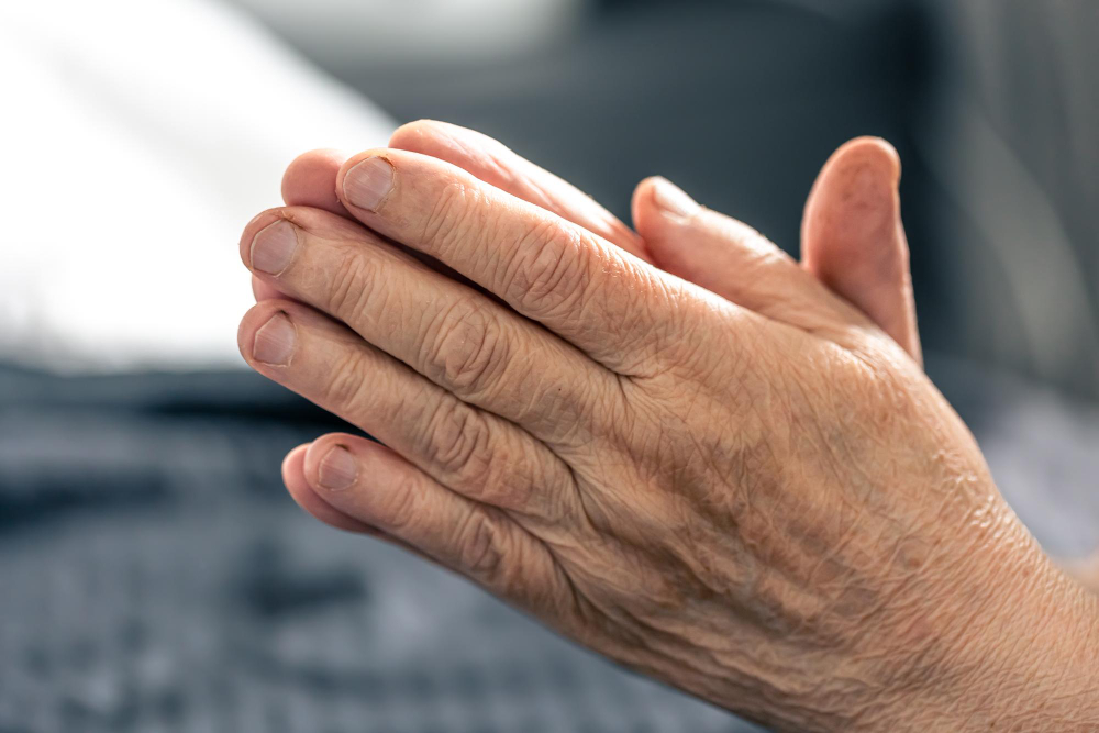
Image by pvpproductions, Freepik, Licence
In the integumentary system, the skin:
- acts as a barrier and protects internal organs
- synthesises Vitamin D, an essential substance that helps build and maintain bones
- allows us to respond to pain, pressure, touch and changes in temperature to keep us safe from injury and infection.
The integumentary system consists of many structures to support the functions of the body.
Select to learn more
Select the items on the image to reveal more information.
Image by macrovector, Freepik, Freepik licence
Important
The subcutaneous layer, also known as the subcutis or superficial fascia, is the deepest layer of the skin and connects the skin with the muscles and bones.
It consists primarily of fat and areolar connective tissue to help the skin stretch and move.
The subcutaneous layer contains adipose tissue which helps to insulate the body, store energy and protect the organs beneath it.
People often use ‘hypodermis’ and ‘subcutaneous’ interchangeably to refer to the same layer of tissue beneath the skin.
Functions of the integumentary system
The integumentary system has many functions including protection, temperature regulation, Vitamin D synthesis, sensation and excretion.
Select to learn more
Select each bar to expand and reveal further information about the integumentary system.
 Watch the video
Watch ‘The science of skin’ to learn more about the integumentary system. Then complete the quiz
Check your understanding
Respond to the questions below and select ‘Check’ to see if you are correct. Select the 'Next' button to move to the next questions, then select 'Check' again for feedback.
Lymphatic system
The lymphatic system plays an important role in immunity by defending the body against pathogens (disease-causing organisms), by filtering, removing and reacting to them. Immunity refers to the state of being resistant or insusceptible to pathogens or infectious diseases.
People with compromised immune systems or inefficient lymphatic systems need special care. They should be kept away from allergens and people with contagious diseases. The lymphatic system forms part of the immune system and is made up of:
- nodes located at various places throughout the body, including in the neck, under the arms, in the abdomen and intestines, near the genitals and near the knees
- capillaries and blood vessels
- the thymus
- the spleen
- red bone marrow.
Structures of the lymphatic system
The lymphatic system collects fluid and particles that have moved into the tissue spaces.
Initially the vessels are very thin with openings to allow fluid and material to enter. The vessels gradually change and the walls become thicker and vein-like with valves to keep the fluid moving in one direction.
Along the pathway, the fluid passes through lymph nodes, which filter the lymphatic fluid. The main cells found in the lymphatic fluid are lymphocytes which are white blood cells dedicated to fighting infection. In the nodes, special cells (lymphocytes and macrophages) collect and destroy harmful matter, such as bacteria. When an infection is present, the lymph nodes become swollen as more of the special cells are required to collect the bacteria.
People with excess fluid in their tissues have a condition called oedema. Their tissue/limbs become swollen, and in severe cases, fluid can leak through the skin.
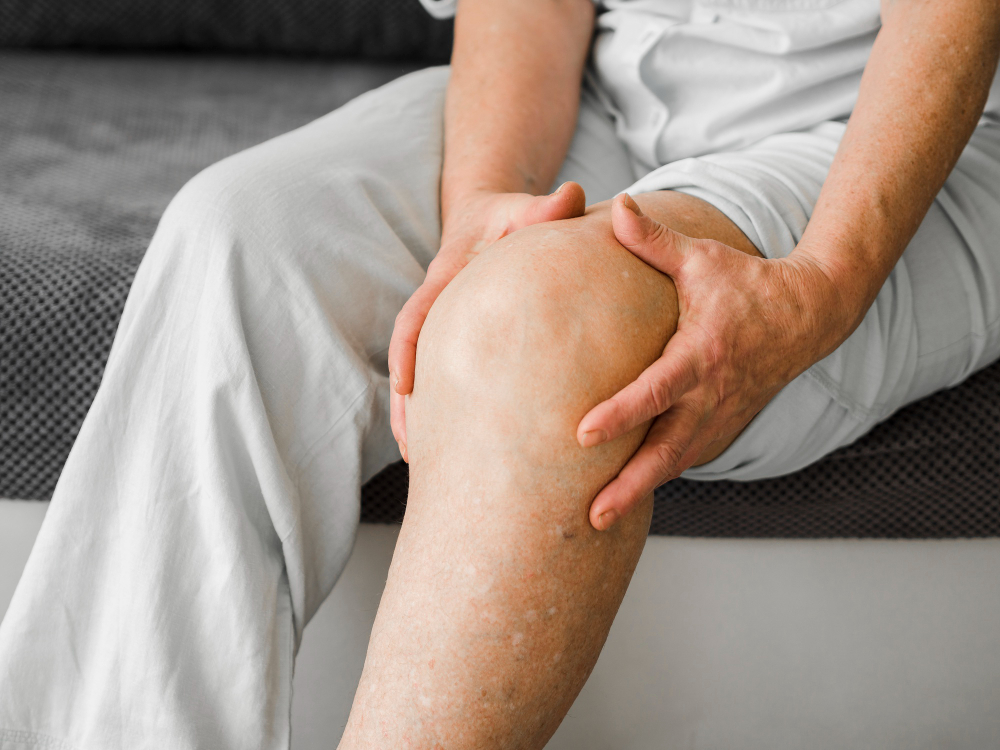
Image by Freepik, Freepik, Licence
Some body organs also form part of the lymphatic system. These organs are the tonsils, adenoids, spleen and thymus. Other lymphatic tissue is found throughout the body.
The lymphatic fluid is eventually returned to the circulatory system, emptying into the large venous vessels of the neck.
Select to learn more
Select each bar to expand and reveal further information about the structures of the lymphatic system.
Location of the lymphatic system
The lymphatic system is similar to the cardiovascular system in that it covers all regions of the body. It consists of glands vessels, nodes, tissues and organs.
When the lymphatic system is working to protect the body from disease, the nodes that consist of lymphatic tissue increase in size and can be felt through the skin.
Lymphoid organs are categorised as primary or secondary organs:
- The primary organs for the immune system are the bone marrow found within all bones of the body and the thymus, which is located in the chest and active before puberty.
- The secondary lymphoid organs include the spleen, lymph nodes, tonsils, appendix and Peyer’s patches. Tonsils are found in the back of the throat, lymph nodes are scattered throughout the body, and the spleen, appendix and Peyer’s patches are located within the abdomen.
 Select to learn more
Select the items on the image to reveal more information about lymphatic system structures throughout the body.
Image by drobotdean, Freepik, Licence
 Watch the video
Watch ‘The Lymphatic System’ to learn more about the lymphatic system. Then complete the quiz
Check your understanding
Respond to the questions below and select ‘Check’ to see if you are correct. Select the 'Next' button to move to the next question, then select 'Check' again for feedback.
Immune system
The immune system’s main function is to fight infection.
The main components of the immune system are specialised white blood cells that help the body overcome infection. These specialised cells are B cells and T cells and are made in the primary lymphoid organs, the bone marrow and the thymus.
A four-stage process is followed by the body’s immune system to protect the body from infection, which includes:
| Step | Description |
|---|---|
| 1. Recognition | Foreign material is identified. |
| 2. Activation and Mobilisation | A signal is sent through the attachment of a T cell to the foreign microorganism or B cell recognition, and white blood cells travel to the site. |
| 3. Regulation | Damage to the body by the immune response is controlled by suppressor T cells. |
| 4. Resolution | This step confines the invading material and eliminates it from the body. The white blood cells then self-destruct and are ingested; however, some are not destroyed and become memory cells. |
The immune system is closely linked to the lymphatic system and the circulatory system.
Secondary lymphoid organs are found throughout the body and consist of the spleen, lymph nodes, tonsils, appendix and Peyer’s patches in the intestines.
Cells within the immune system that assist with the fight against infection or foreign material include:
- killer cells that attack infection
- suppressor cells that stop the immune response
- helper cells that assist to make antibodies.
Components of the immune system
The components of the immune system are white blood cells and the lymphoid organs. The white blood cells that fight infection can move freely throughout the whole body and travel within the lymph fluid and the blood.
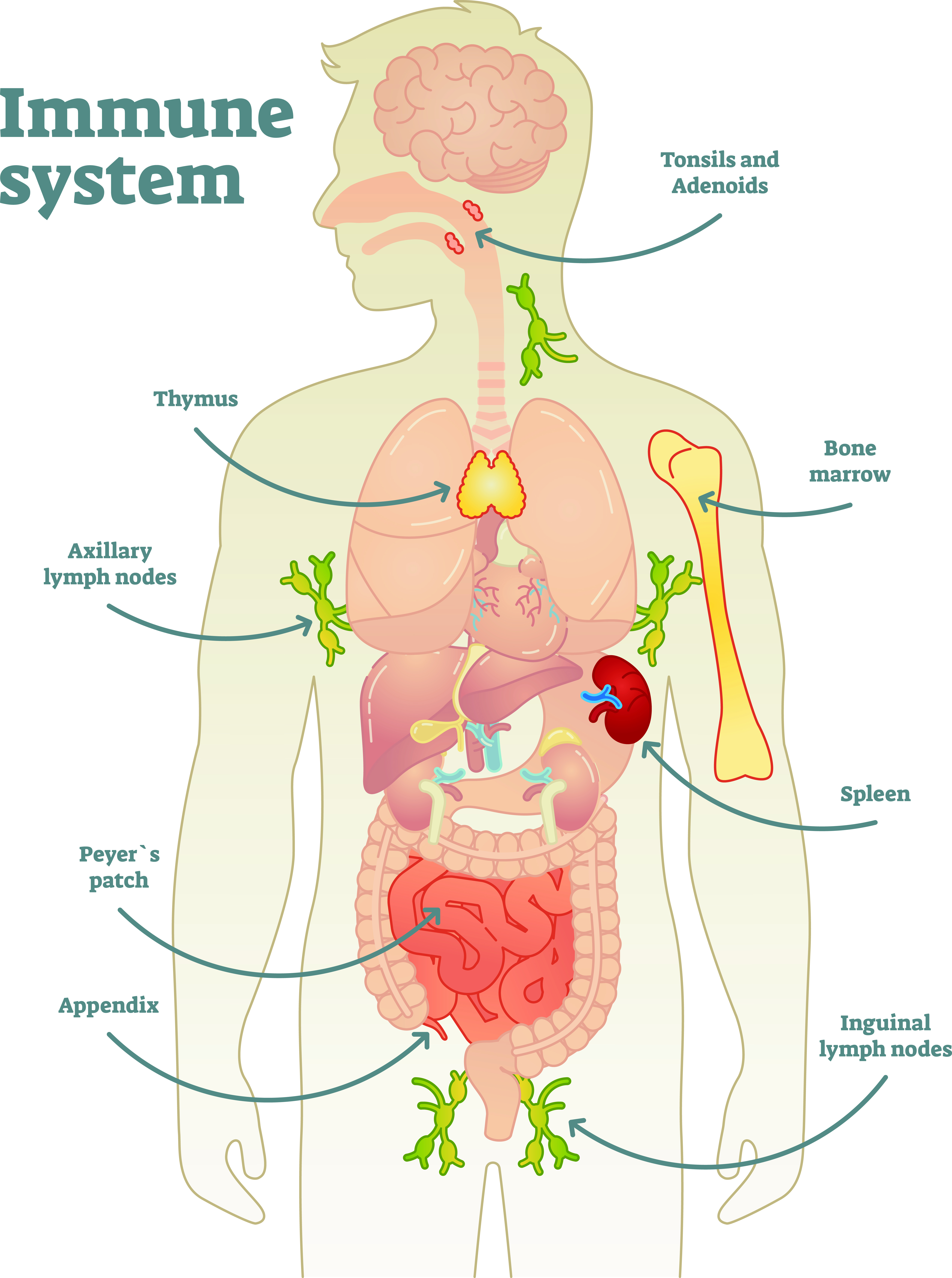
Image by VectorMine, Shutterstock, Shutterstock licence
Select to learn more
Select each bar to expand and reveal further information about the components of the immune system.
The immune system at work
The immune system defends the body against foreign or dangerous invaders such as:
- microorganisms
- parasites
- cancer cells
- foreign tissues and organs (transplants).
It is able to detect what belongs to the body and what doesn’t and will initiate an immediate response to the threat which is known as an immune response. The immune response can be an increase in production of white blood cells by the nodes and organs, movement of the white blood cells to a particular location of the body, and/or a fever.
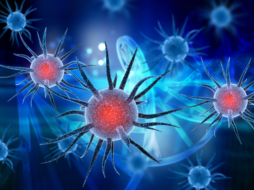
Image by kjpargeter, Freepik, Licence
Our immune system helps protect us when the first line of defence of the body is overcome. This happens, for instance, when we cut ourselves and microbes enter through this opening. Microbes may be viruses, bacteria and parasites. Viruses tend to be airborne and usually enter the body through the respiratory system.
A chemical message is sent to the white blood cells by the damaged cells of the body. The white blood cells move to the affected area and engulf the invading microorganisms. A secretion (pus) is produced, which is a mixture of lymph fluid, white blood cells and dead microorganisms.
Fever can be part of the immune response and is the body raising its temperature so it becomes a less favourable host. Fever also supports the immune response.
The body also tries to organise second lines of defence against the invading microorganisms. Special lymphocytes (B cells) try to identify if they have been in contact with the microorganism previously. If they have, they can generate the antibodies, made from previous exposure, to overcome the microorganisms.
| Defence Level | Description |
|---|---|
| Immune system first line of defence |
Physical or mechanical barriers to infection:
|
| Immune system second line of defence |
|
Watch and answer
Watch ‘How does your immune system work?’ to learn more about the immune system and answer the question as it appears.
Nervous system
The nervous system is responsible for communicating information received by the senses to the brain, processing this information and communicating the response to the muscles of the body.
The nervous system is divided into two parts:
- The central nervous system is made up of the brain and spinal cord. These structures are protected by bone – the skull and vertebral bones of the spine – and the cerebrospinal fluid that cushions the brain and spinal cord.
- The peripheral nervous system is made up of sensory neurons, ganglia and nerves that connect the rest of the body to the spinal cord. There are 12 cranial nerves and 31 spinal nerves that are covered by a special insulating tissue called the myelin sheath. It is these nerves that communicate information to and from the brain.
Image by kjpargeter, Freepik, Licence
The two aspects of the nervous system enable communication within the body with respect to the external and internal environment. The way in which this communication takes place is through the transmission of signals fired by the neurons and conducted along the nerve pathways of the body.
Components of the nervous system
Each component of the nervous system comprises many parts. Information from sensory organs such as the skin, eye and ear are transmitted along various pathways until it reaches the brain where information processing takes place.
For example:
- the eye takes in information from the environment
- light enters through the pupil and travels through the lens to the optic disc
- an impulse is generated and is transmitted along the optic nerve to the brain for processing.
Select to learn more
Select each bar to expand and reveal further information about the components of the nervous system.
Functions of the nervous system
The brain processes the information from the nerves. Sensory neurons transmit messages to the brain and motor neurons
take messages from the brain to the body.
Nerves are made up of many neurons, which transmit or relay the messages to each other along a pathway called a nerve. Each nerve is made up of a number of neurones. The message is transmitted via electrical impulses along the axon and via a chemical messenger at the space between each neuron (the synapse). The first component of the neuron is the dendrite, which captures the chemical message and transfers it to the cell body and then through the axon.
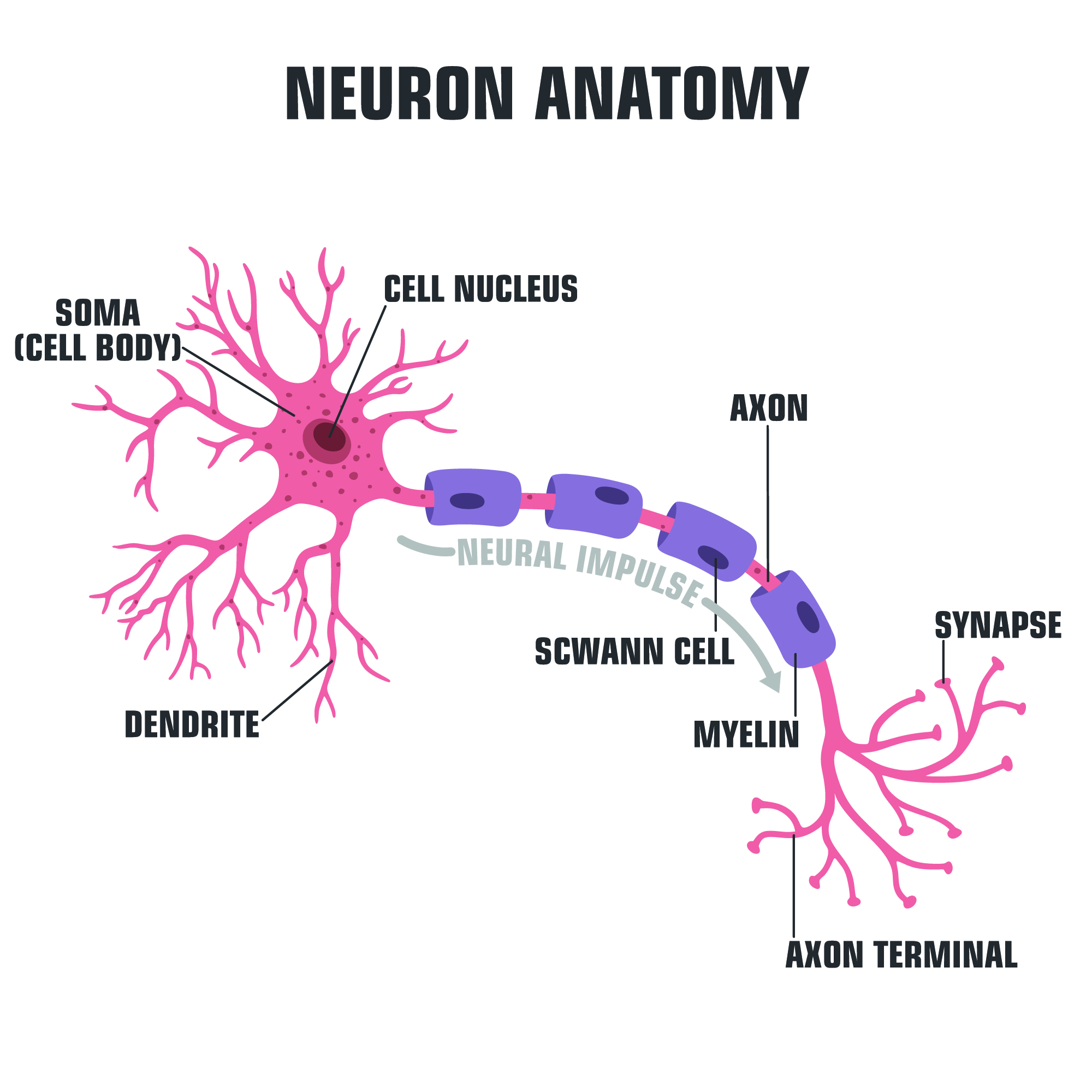
Image by ShadeDesign, Shutterstock, Shutterstock licence
The nervous system also has a specialised pathway known as a reflex arc to protect the body under certain potentially dangerous circumstances. It is a pathway of impulse transmission that travels from the sensory nerve to the spinal cord and directly out again via a motor nerve to muscles that can act on the danger. At the same time the message is conveyed to the brain.
An example of a reflex arc is when you blink your eye when a puff of wind hits it, or take your hand away from a hot stove even before you register pain. This is a way the body protects itself from harm.
| Nerve | Description |
|---|---|
| The optic nerve |
The optic nerve is located in the back of the eye. It is the second cranial nerve. There is a nerve to both the right and left eye. These nerve fibres separate at an X-shaped space in front of the brain. At this point of the optic nerve, the part of the nerve close to the nose crosses over. |
| The auditory nerve |
The auditory nerve transfers auditory information from the cochlea to the brain. It is the eighth cranial nerve. The function of the cochlear nerve is to gather auditory data from the environment. |
 Watch the video
Watch ‘The nervous system in 9 minutes’ to learn more about the nervous system. Then complete the quiz.
Check your understanding
Respond to the questions below and select ‘Check’ to see if you are correct. Select the 'Next' button to move to the next questions, then select 'Check' again for feedback.
Special senses
The special senses are part of the nervous system providing vision, hearing, smell, taste and equilibrium. These senses are used to detect changes in external stimuli so that one can react and respond appropriately. Examples of external stimuli include fumes, temperature, sounds and changes in movement.
The nose, ears, eyes and mouth are responsible for sensing external stimuli. This stimulus (the smell, sound, image or taste) is interpreted by the brain. Damage to the brain or certain brain disorders may alter the way the smell, taste, aural or visual information is perceived. Sensation and perception are interlinked, but they are distinct processes.

Image by user18526052, Freepik, Licence
Smell
The sense of smell is the ability to detect odours. Smell is used to detect danger such as fumes and gases. A person with a diminished sense of smell cannot taste food and may not notice their own odours, leading to social embarrassment.
Smell is only one of the nose’s functions. Noses also warm and filter the air we inhale and prevent foreign bodies from entering the respiratory system.
Let’s explore some terminology that is related to the sense of smell.
| Term | Description |
|---|---|
| Cilia |
The tiny hairs in the nose. |
| Nostrils |
The two openings of the nose. |
| Olfactory |
Relating to the nose and sense of smell. |
| Septum |
The wall between the nostrils. |
 Watch the video
Watch ‘How do we smell?’ to learn more about the olfactory sense.
Make notes in your notebook or digital device to refer to later.
Check your understanding
Fill in the blanks by dragging the words into the correct place then select ‘Check’ to see if you are correct.
Taste
The sense of taste is the ability to detect the flavours of foods and other substances. The tongue has a number of receptors (taste buds) that detect whether food is sweet, sour, salty, bitter or umami.
These receptors allow people to detect whether food is off, which is important to one’s safety. Taste buds also enhance our enjoyment of food. As people age, these receptors become less effective and can affect an older person’s enjoyment of food.
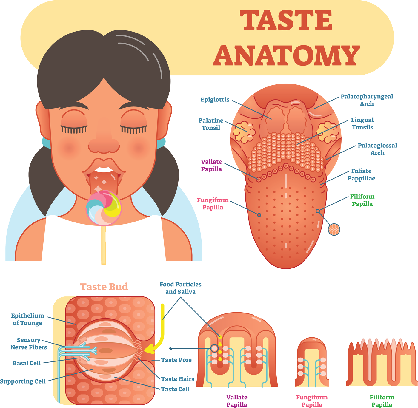
Image by VectorMine, Shutterstock, Shutterstock licence
Let’s explore some terminology that is related to the sense of taste.
| Term | Description |
|---|---|
| Oral |
By mouth. |
| Xerostomia |
Dry mouth. |
| Saliva |
A fluid secreted into the mouth that helps break down food. |
Sight
The eyes provide the sensory stimuli that allow for vision, which lets people see what is around or near them.
The outer tissue layer of the eye is called the cornea. It is transparent and very sensitive.
Light enters the eye through the pupil and is refracted by the lens through the vitreous gel (the liquid in our eyes). A signal is then sent to the brain from the retina via the optic nerve.
People’s ability to see can be reduced as they grow older. Vision impairment can also arise because of disease or illness.
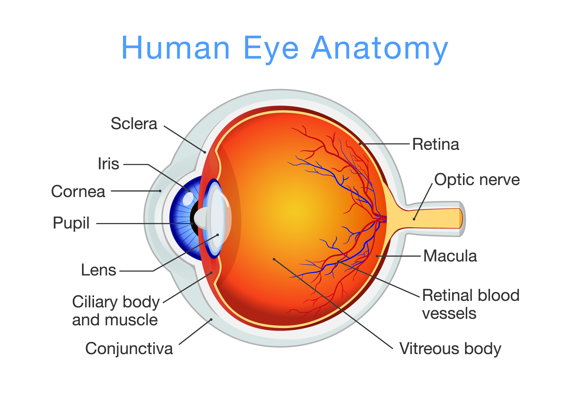
Image by solar22, Shutterstock, Shutterstock licence
| Optical Structure | Function |
|---|---|
| Cornea | The cornea is the clear outer part of the eye’s focusing system located at the front of the eye. |
| Pupil | The pupil is the opening at the centre of the iris. The iris adjusts the size of the pupil and controls the amount of light that can enter the eye. |
| Iris | The iris is the coloured part of the eye that regulates the amount of light entering through the pupil. |
| Macula | The macula is the small, sensitive area of the retina that gives central vision. It is located in the centre of the retina. |
| Vitreous gel | The vitreous gel is a transparent, colourless mass that fills the rear two-thirds of the eyeball, between the lens and the retina. |
| Lens | The lens is a clear part of the eye behind the iris that helps to focus light, or an image, on the retina. |
| Retina | The retina is the light-sensitive tissue at the back of the eye. The retina converts light into electrical impulses that are sent to the brain through the optic nerve. |
| Optic nerve | The optic nerve is the largest sensory nerve of the eye. It carries impulses from the retina to the brain. |
Source: Eye Anatomy Handout
 Watch the video
Watch ‘How the eye works animation’ to learn more about how we see. Then complete the quiz.
Check your understanding
Respond to the question below and select ‘Check’ to see if you are correct.
Equilibrium
Equilibrium means balance. The inner ear plays a role in maintaining bodily equilibrium. The inner ear contains fluid which, along with our vision, helps us determine whether we are moving, stationary, upright or lying down.
Balance disorders can cause vertigo, dizziness and nausea. Balance is particularly important for older people who are more susceptible to breaks and fractures when falling.

Image by Freepik, Freepik, Licence
Hearing
The ears are responsible for detecting, amplifying, transducing and interpreting sound waves, allowing us to hear and understand the world around us. They are also responsible for maintaining balance.
The ears capture sound waves and transform them into something our brains can understand.
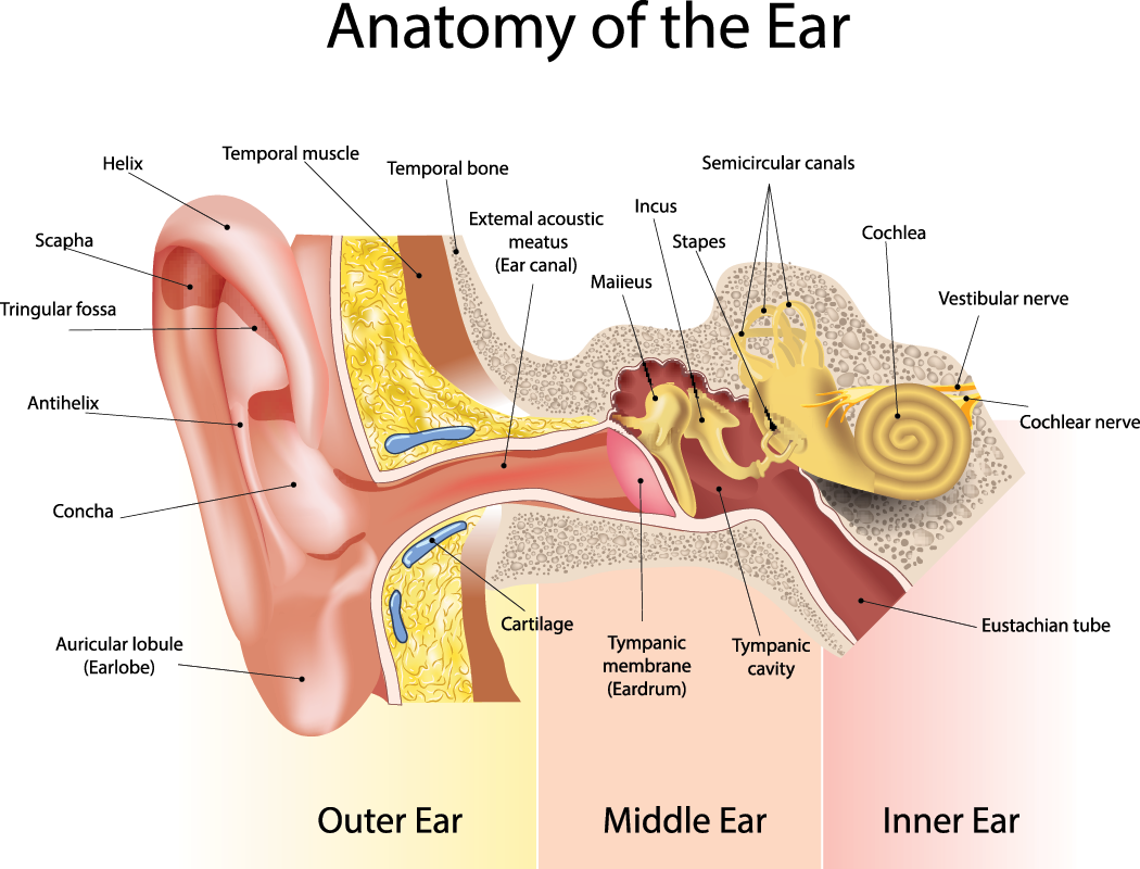
Image by SVETLANA VERBINSKAYA, Shutterstock, Shutterstock licence
The process starts with the outer ear, where the pinna collects sound and directs it through the ear canal to the eardrum. As the eardrum vibrates from the incoming sound, it sets off a chain reaction with three tiny bones in the middle ear – the hammer, anvil and stirrup – which amplify these vibrations.
These amplified vibrations then enter the cochlea, a fluid-filled structure in the inner ear. Inside the cochlea,
thousands of tiny hair cells convert these vibrations into electrical signals, which travel through the auditory
nerve to the brain. Once in the brain, these signals are interpreted, allowing us to perceive and understand the
sounds in our environment.
Let’s explore some terminology that is related to the sense of hearing.
| Term | Description |
|---|---|
| Aural | By ear. |
| Audio | Relating to sound. |
| Audiologist | A specialist ear doctor. |
 Watch the video
Watch ‘The science of hearing’ to learn more about how we hear. Then complete the quiz.
Check your understanding
Respond to the questions below and select ‘Check’ to see if you are correct. Select the 'Next' button to move to the next question, then select 'Check' again for feedback.
Now that you've explored each system, complete the following quiz.
Check your understanding
Respond to the questions below and select ‘Check’ to see if you are correct. Select the 'Next' button to move to the next question, then select 'Check' again for feedback.
Background Colour
Font Face
Font Kerning
Font Size
Image Visibility
Letter Spacing
Line Height
Link Highlight
Text Colour